An MRI (magnetic resonance imaging) examination is a test that generates clear pictures of the edifices inside your body using a huge magnet, radio waves, and a computer. Healthcare providers use MRIs to assess, identify and monitor several diverse therapeutic circumstances.
What is an MRI Examination?
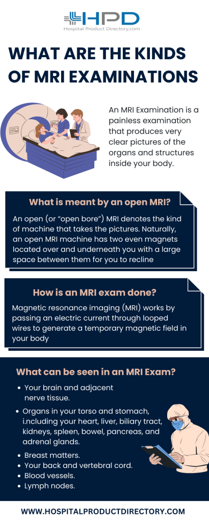
An MRI (magnetic resonance imaging) examination is a painless examination that produces very clear pictures of the organs and structures inside your body. The MRI machine made by MRI Machine Manufacturers uses a huge magnet, radio waves, and a computer to produce these comprehensive pictures. Because MRI doesn’t use X-rays or other radioactivity, it’s the imaging examination of choice when people will need recurrent imaging for analysis or treatment monitoring, particularly of their brain.
What is meant by an open MRI?
An open (or “open bore”) MRI denotes the kind of machine that takes the pictures. Naturally, an open MRI machine has two even magnets located over and underneath you with a large space between them for you to recline. This permits open space on two sides and eases much of the claustrophobia many people experience with closed-bore MRI machines.

Though, open MRIs don’t take as strong pictures as closed-bore MRI machines. Closed-bore MRI machines have a circle of magnets that forms an open hole or cylinder in the middle where you’d recline to get the pictures. Closed-bore MRIs are slender with constricted head-to-ceiling space. This can reason nervousness and discomfort for some people, but these MRI machines take the best quality pictures.
If you’re anxious about your MRI examination or have a fear of closed spaces, talk to your healthcare provider. If desired, your provider will deliberate choices for tranquilizers (drugs to make you feel relaxed) or even anesthesia if essential.
Why is an MRI with contrast done?
Some MRI examinations use an inoculation of contrast material. The contrast agent comprises gadolinium, which is a sporadic earth metal. When this material is current in your body, it modifies the magnetic properties of neighboring water molecules, which improves the quality of the pictures. This advances the sensitivity and specificity of the diagnostic pictures.
Contrast material improves the discernibility of the following:
- Cancers.
- Irritation.
- Contagion.
- Bloodstream to confident organs.
- Blood vessels.
If your MRI needs contrast material, a healthcare provider will implant an intravenous catheter (IV line) into a vein in your hand or arm. They’ll use this IV to vaccinate the contrast solid.
Contrast ingredients are safe medications. Side effects fluctuating from slight to severe do arise, but severe reactions are very rare.
How are MRI examinations different from CT scans?
Magnetic resonance imaging (MRI) uses magnets, radio waves, and a computer to generate pictures of the inside of your body, whereas computed tomography (CT) uses X-rays and computers.
Healthcare providers often favor using MRI examinations instead of CT scan to look at the non-bony parts or soft tissues inside your body. MRI images are also harmless since they don’t use the harmful ionizing radiation of X-rays. MRI examinations also take much clearer images of your brain, spinal cord, nerves, muscles, ligaments, and tendons than steady X-rays machines and CT examinations.
Though, not everybody can undergo an MRI. The magnetic field of MRI can dislocate metal grafts or affect the purpose of devices such as pacemakers and insulin pumps. If this is the circumstance, a CT examination is the next best option. MRI examination is typically costlier than X-ray imaging or CT scanning.
What can be seen in an MRI Exam?
Magnetic resonance imaging (MRI) creates thorough pictures of the inside of your body. Healthcare providers can “look at” and assess numerous different structures inside your body using MRI, including:
- Your brain and adjacent nerve tissue.
- Organs in your torso and stomach, including your heart, liver, biliary tract, kidneys, spleen, bowel, pancreas, and adrenal glands.
- Breast matters.
- Your back and vertebral cord.
- Pelvic structures, including your bladder and generative organs (uterus and ovaries in persons allocated female at birth and the prostate gland in people allocated male at birth).
- Blood vessels.
- Lymph nodes.
When will a patient require an MRI?
Healthcare providers use magnetic resonance imaging (MRI) to assist in identifying or monitoring the treatment for many diverse conditions. There are also diverse kinds of MRIs done on equipment supplied by MRI machine Suppliers founded on which part of your body your provider wants to examine
How safe is an MRI Exam?
An MRI examination is usually safe and poses almost no danger to the average person when suitable safety rules are followed. The strong magnetic field the MRI machines produce is not damaging to you, but it may cause entrenched medical devices to break down or misrepresent the pictures. There’s a very small danger of an allergic reaction if your MRI needs the use of contrast material. These responses are usually slight and controllable by medicine. If you have an allergic reaction, a healthcare provider will be obtainable for instant help.
Healthcare providers usually don’t perform gadolinium contrast-enhanced MRIs on expectant people due to unidentified risks to the developing baby unless it’s essential.
Who must not get an MRI Exam?
In most circumstances, an MRI exam is harmless for people with metal grafts, except for a few sorts. Unless the device you have is approved as MRI-safe, you may not be able to have an MRI. These machines may comprise:
- Metal joint prostheses.
- Some cochlear grafts.
- Some kinds of pins are used for brain aneurysms.
- Some kinds of metal loops are placed within blood vessels.
- If your healthcare provider endorses an MRI examination, they’ll ask comprehensive questions about your medical history and any medical devices or grafts you may have in or on your body.
How is an MRI exam done?
Magnetic resonance imaging (MRI) works by passing an electric current through looped wires to generate a temporary magnetic field in your body. A transmitter/receiver in the MRI machine bought from an MRI machine Dealers then directs and receives radio waves. The computer then uses these signals to make digital pictures of the perused part of your body.
How does one get ready for an MRI?
The magnetic resonance imaging (MRI) scanner uses strong magnets and radio wave signals that can reason the warming or conceivable movement of some metallic objects in your body. This could result in fitness and safety issues. It could also cause some fixed electronic medical devices to break down.
If you have metal-containing substances or entrenched medical devices in your body, your healthcare provider needs to know about them before your MRI examination. Certain entrenched objects may need additional scheduling preparations and special orders. Other items don’t need special orders but may need an X-ray to check on the exact position of the object before your examination.
Leave all jewelry and other fittings at home or eliminate them before your MRI examination. Metal and electronic items aren’t permitted in the examination room because they can hinder the magnetic field of the MRI unit, cause blisters or become harmful projectiles.
What is the period of an MRI Exam?
Contingent on the kind of examination and the equipment used, the complete examination usually takes 30 to 50 minutes to complete. Your healthcare provider will be able to give you a more meticulous time assortment based on the exact reason for your examination.
What must I suppose during an MRI?
Most MRI examinations are effortless, but some people find it painful to remain still for 30 minutes or longer. Others may know nervousness from the closed-in space while in the MRI machine. The apparatus can also be loud.
In some instances, your MRI may require disparity. If this applies to you, a provider will give you an IV inoculation of contrast material before you experience the MRI. The IV pointer may cause some uneasiness but this won’t last long. You may have some staining later. Some people know a provisional metallic taste in their mouth after the contrast inoculation.
Does MRI contrast the reason for any side effects?
On very infrequent events, some people who have contrast material for their MRI knowledge have side effects, including:
Biliousness.
Headache.
Aching at the place of the inoculation.
It’s very infrequent to knowledge rash, prickly eyes, or other signs of an allergic response to the contrast material. If you have sensitive indications, tell the technologist. A healthcare provider will be obtainable to offer instant medical care.
Nephrogenic systemic fibrosis (NSF), which causes congealing of your skin, organs, and other tissues, is a rare problem in people with kidney illness that experience an MRI with contrast material. Because of this, people with acute kidney illness may not be able to have gadolinium-based contrast material for their MRI.
There’s an indication that tiny suggestions of gadolinium may stay in diverse organs of your body after contrast-enhanced MRI. While there are no recognized undesirable effects from this, your provider may take gadolinium holding into account when choosing a contrast agent.
When will a patient get to know the outcome of the MRI?
After the MRI examination, a doctor will examine the pictures. The radiologist will guide an initial report to your primary healthcare provider, who will apportion the consequences with you. You may want a continuation examination. If so, your doctor will explain why.
