What is a Neuro Microscope?
Various illnesses, including cancer, need surgery as a prime treatment method. One key issue for surgeons to operate precisely is a clear visualization of the anatomical structures. Though, this has never been relaxed. On the one hand, some bodily structures are very small, fluctuating from millimeters to microns, and they might have close proximity to other organs or tissue. A clear opinion of these constructions needs a resolution well beyond that of human eyes. On the other hand, the lack of lighting in narrow hollows and deep channels, which are very shared in neurosurgery, results in a dim conception of shadows. Poor conception may lead to inappropriate operation on anatomical edifices or a nearby organ, which will affect the operating outcome, decrease organ preservation, or even cause life-threatening consequences. Therefore, adequate exaggeration and proper lighting are vital for the success of the surgery.
With abundant advantages such as clear and cheerful visualization, easy documentation and adaptation, constancy, maneuverability, and enhanced ergonomics, Neuro Microscopes made by Neuro Microscope Manufacturers have been applied in various kinds of surgeries, including neuro and spine surgery. For instance, they have been used for brain cancer resection, aneurysm surgery, and head and neck cancer resection. For different applications, microscopes are adapted into slightly diverse optical outlines and equipped with specific imaging modalities. The end-users of Neuro Microscope comprise hospitals, dental clinics, other outpatient settings, and some research organizations.
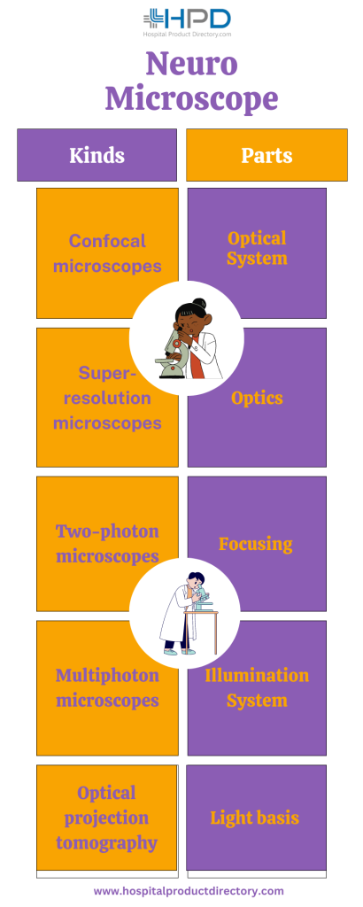
There are numerous kinds of neuro microscopes available, including:
- Confocal microscopes: These use laser light to selectively irradiate and image individual planes of a sample, producing high-resolution pictures of thick tissue samples.
- Two-photon microscopes: Alike to confocal microscopes, two-photon microscopes use lasers to infiltrate deep into the tissue, but they can also be used to complete in-vivo imaging.
- Multiphoton microscopes: These are alike to two-photon microscopes but can notice numerous photons produced from the specimen, enabling earlier and more efficient imaging.
- Electron microscopes: These use a ray of electrons to yield high-resolution pictures of ultrastructural particulars, counting the internal structure of cells and the fine details of synapses.
- Optical projection tomography (OPT): This is a kind of 3D imaging technique that uses a light source and a digital camera to produce high-resolution pictures of a specimen.
- Super-resolution microscopes: These use dedicated imaging methods to realize resolution beyond the deflection limit of light, allowing the conception of subcellular structures.
Parts of a Neuro Microscope
The acceptance and modularization of cutting-edge technologies for image-guided surgery have been actively appraised in recent decades. On the one hand, the intraoperative imaging modalities have been assessed with Neuro Microscopes bought by Neuro Microscope Manufacturers to deliver real-time diagnostic information. The imaging modalities employ certain assets of human tissue and disclose information that is beyond what human eyes can see, even the deeper edifices beneath the tissue surface. To apply these imaging modalities, certain scheme versions have been done for the microscope. The objective of the system version is to allow and incapacitate these imaging purposes easily without interjecting the surgical workflow or decreasing the presentation of the microscope.
On the other hand, AR has been playing an imperative role in new-generation microscopes, particularly with the development of minimally invasive surgery. It supports surgeons relating the preoperative two-dimensional (2D) pictures with the real 3D surgical site intraoperatively for navigation. AR can work with various image modalities, either preoperative or intraoperative, and overlap the pictures onto the surgical site so the surgeons do not need to switch their sight between the operating site and pictures. In addition, the overlap of pictures discloses not only the 3D model but also the bodily structures beneath the patient’s skin. With proper system type, accurate calibration and registration, and suitable visualization methods, AR could greatly support the clinic for surgery.

Optical System: The optical system of the microscope is the main cause of the imaging quality that a system can attain. It is essentially binocular (with eyepieces on top) with a close-up lens, specifically the optical apparatuses counting the objective lens and the exaggeration changer (or zoom changer). The focal span of the objective lens fully regulates the value of working distance, which is the distance from the objective lens to the point of emphasis of the optical system. The zoom changer is either a sequence of lenses moving in and out of the watching axis or a system that varies the relative positions of lens elements.
Optics : The plan of optics is vivacious to the image quality of a Neuro Microscope. The deviation is an intrinsic property of optical systems, and it reasons the distortion or alteration of pictures, which is opposed to the desire for a clear view. Monochromatic deviations such as spherical deviation, coma, and astigmatism can be modified but typically only for one color.
Focusing: Concentrating is vital for a clear view. Surgeons would want the operating site to be an emphasis throughout the surgery. Though, the figure of organs or the deep hollows makes it unbearable for the whole operating site to be flawlessly on the focal plane. Depth of emphasis, in other words, depth of field (DOF), is a term that designates the part in front of and behind the point of perfect focus where the sharp focus is upheld. It is contingent on many issues, counting but not restricted to the quality of an optical plan, the scope of the objective lens orifice relative to the focal span of the objective lens, and the exaggeration of the object, and it is mutual of the resolution. A good Neuro Microscope must have an acceptable depth of focus without forfeiting too much resolution to keep the scene shrill.
Illumination System: Brightness is another key issue besides the optical system for the imaging quality of a microscope. Efficacious surgical lighting has four key factors, specifically luminance, shadow management, volume of light, and heat. A positive view of the whole operating site during the surgery is always desired. The innovative illuminator in the original Neuro Microscope was an autonomous bulb outwardly affixed on the side of the microscope. Light conveyed to the operating site likely generates shadows, and thus illumination of deep cavities was hardly possible. Modern microscopes have espoused high-power light sources with stable light strength and close-to-sunlight color temperature.
Despite plentiful returns of Neuro Microscope brilliance, it is still worth noting that many up-to-date neuro and spine surgical microscopes use light bases of the highest strength to deliver the best illumination and clearness for human eyes irrespective of magnification and working distance. Though, the high power can damage the fundamental tissue. Though Neuro Microscope Manufacturers deliver safety cautions of possible damage, specific settings of the lighting are not regulated.
Light basis: Except for the out-of-date glowing bulbs used in old Neuro Microscopes, there are mainly three kinds of light causes, i.e., xenon lamp, halogen lamp, and LED. LEDs can provide lighting in the visible wavelength array with good illumination, good constancy, longer life, less power consumption, and extremely low heat. Though, LED as a surgical light basis also has drawbacks: the higher color temperature and thinner wavelength array make the light not as close to sunlight; its band is inadequate for fluorescence-guided applications, particularly ICG imaging, where an excitation light in the NIR array is required; furthermore, it is not easy to substitute.
Illumination arrangement : The tissue exterior being viewed under a Neuro Microscope during operation is usually damp and highly reflective. The light that originates from an angle can be easily reproduced away and cause a dark view. Coaxial lighting is the answer to this situation. Different from lateral lighting where light comes from the side, coaxial lighting matches the optical axes of lighting and conception (lens). Lighting from the light basis that locates on the side is unfocussed and anticipated almost parallel to the axis of the lens. Consequently, light vertically illumines the tissue surface and is reproduced directly to the lens, not having much loss. Coaxial lighting decreases the width of the floodlit area, furthermore, it can be directed into thin and deep hollows, which is helpful for neurosurgery.
