Mammography, also recognized as mastography is the way toward using low-vitality X-beams for the most part about 30 kVp to examine the human breast for analysis and screening.
What is the main purpose of Mammography?
The purpose of mammography is the early detection of breast cancer, normally through the detection of trademark masses or microcalcifications.
Why are Mammograms done?
Mammography is X-ray imaging of distinct breasts envisioned to recognize tumors and different disparities from the norm. Mammography can be used either for screening or for analytical purposes in evaluating a breast irregularity:
Screening mammography: Screening mammography is used to recognize breast changes in ladies who have no signs or side effects or new breast differences from the norm. The intention is to identify cancer before medical signs are noticeable.
Analytical mammography: Analytical mammography is used to research breast cancer changes, for instance, another breast anomaly, breast nuisance, an uncommon skin advent, nipple thickening, or areola release.

It’s likewise
used to evaluate asymmetrical detections on a screening mammogram. An analytic
mammogram done on equipment made by Mammography Machine Manufacturers comprises
extra mammogram images.
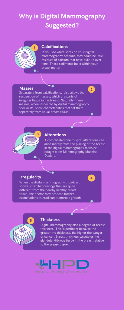
Why is digital mammography suggested?
When your
doctor endorses that you get a mammogram, they expect to obtain certain
findings and remove certain risks. When the mammogram is being read by the radiologist,
they will be on the watch for any irregularities that may designate a disease
or injury. Naturally, the radiologist looking at your digital mammography
account will be watching for:
● Calcifications: If you see white spots
on your digital mammography account, they could be little residues of calcium
that have built up over time. These sediments build within your breast matter.
These might be of two types:
●
Macrocalcification: These are comparatively
large sediments in the breast tissue that may ascend due to the aging of
arteries or irritation. They might also be the consequence of some old
injuries. Overall, macrocalcification spots are not examined further because
they do not designate a cancerous disorder. In elder females, that is, above
50, these macro-calcifications are quite a shared marvel.
●
Microcalcification: These are lesser white
spots that can be noticed by digital mammography experts to screen for breast
cancer. While microcalcifications do not settle cancerous evolution, they are
taken more gravely than macrocalcifications. Knowledgeable digital mammography
experts study the form and location of these spots and their nearness to tissue
masses to gauge the possible danger of cancer. In precise cases, contingent on
the pattern or appearance of the microcalcification spots, the doctor may
endorse a biopsy to recognize if the growth is cancerous.
● Masses: Separately from calcifications,
digital mammography is done on equipment supplied by Mammography Machine
suppliers also allows the recognition of masses, which are
parts of irregular tissue in the breast. Naturally, these masses, when
inspected by digital mammography specialists, show characteristics that set
them separately from usual breast tissue. While masses can be just
liquid-filled pockets or rock-hard tumors, they can also be tumorous, which is
when digital mammography transmission helps in early identification and
analysis.
● Alterations: A complicated one to spot,
alterations can arise merely from the placing of the breast in the digital
mammography machine bought from Mammography Machine Dealers.
Such alterations also ascend from some preceding injuries to the breast area or
from previous procedures that were completed. Though this is also a likely sign
of cancer, so the digital mammography transmission again helps in early
discovery and analysis.
● Irregularity: When the digital
mammography broadcast shows up white coverings that are quite different from
the nearby healthy breast tissue, the doctor may propose further examinations to
eradicate tumorous growth. Numerous types of asymmetries may be produced for
different reasons.
● Thickness: Digital mammography also a
degree of breast thickness. This is pertinent because the greater the
thickness, the higher the danger of cancer. Breast thickness calculates the
glandular/fibrous tissue in the breast relative to the greasy tissue. Higher
breast thickness is flawlessly normal, but it does place one at a slightly
raised danger of cancer, which means that more recurrent checks might be necessary.
If your
clinician has asked you to get digital mammography done sporadically, then the
reports will usually be associated with previous ones. The radiologist will try
to assess if any changes can be seen since the last time you had a mammogram
completed. Their study can display any new growths or further changes in an
existing mass or spot that was evident in previous reports.
What is full turf digital mammography?
Before we get
into what a full turf digital mammography or FFDM is, let’s comprehend what a
mammogram is. Merely put, a mammogram is nothing but a low-dosage X-ray that is
understood by radiologists to recognize any changes in the tissue in the breast
region. Digital mammography is also named full turf digital mammography, and
this is a process where instead of the traditional X-ray film, you have
solid-state sensors transferring the X- rays into electrical signals. These
signals reconstruct pictures of the breast, which can be further considered on
a computer screen. You can also get a picture yield on special film. Far ahead,
Computer Aided Design or CAD can ease the examination of breast tissues more
precisely and professionally using digital mammography output. The CAD system
sketches areas of anxiety for the doctor to further study, thus plummeting the
human error factor.
What
is 3D digital mammography?
Also recognized as Tomosynthesis, 3D
digital mammography is an improvement in digital mammography that gives 2D and
3D yields. This kind of yield makes it easier to spot irregularities that may
be early tumors or have the potential to turn into growth. In a 3D digital
mammography, the pictures of the breast are given in 1-millimeter thick
sections so the radiologist can study the complete breast in detail, missing
nothing.
How does one prepare for a digital
mammography appointment?
● Delicate breasts may make digital
mammography a more painful procedure than it has to be. Select a time of the
month when your breasts are not Inflamed and book your appointment then.
Typically, this is best after your periods.
● The radiologist will ask for
preceding mammograms to do a proportional examination, so take these reports
and pictures with you.
● Do not use any kind of balm, cream,
fragrance, or even talc under your arms or over your breasts when you go for
your appointment. Evade sprays and antiperspirants too. These can misrepresent
the mammogram.
● You will be requested to eliminate
any jewelry you are wearing above your waist, such as chains, so, if you can,
leave them at home.
● You will be given a robe at the
testing facility that you must wear instead of the clothing that covers your
upper half.
● The operator will ask you to place
yourself before the digital mammography machine in such a way that your breasts
can be positioned on the given platform one at a time.
● A plastic plate that is static
above is pushed down upon your breast to crush it against the platform so the
tissues are spread out and can be taken obviously by the digital mammography
machine. This can reason distress or even pain. If it is too sore, tell the
operator.
● The digital mammography machine
transfers from side to side to get pictures from all sides. To ease the
imaging, you will be requested to hold your breath for a few moments and keep
still.
● The procedure may be replicated
until pictures have been taken from all routes and then the same is done for
the other breast.
Naturally, digital mammography takes
about 20 minutes to complete. Notify the technician if you have breast grafts
or if you are breastfeeding. If you can’t stand motionless for more than a few
minutes, do let the specialist know beforehand so they can organize a cane or
some other support for the period.
